ORTODONTIA CONTEMPORÂNEA: junho 2018

quinta-feira, 28 de junho de 2018

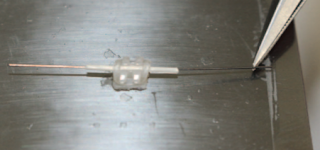
Propriedades mecânicas dos fios ortodônticos cobertos com um tubo de cetona poliéster-éster


Neste artigo de 2018, publicado na Angle Orthodontist, pelos autores Nobukazu Shirakawa; Toshio Iwata; Shinjiro Miyake; Takero Otuka; So Koizumi; Toshitugu Kawata da Division of Orthodontics, Department of Oral Interdisciplinary Medicine (OIM), Graduate School of Dentistry, Kanagawa Dental University, Yokosuka, Kanagawa, Japan. Mostra um estudo laboratorial realizado com arcos ordonticos recobertos com material estetico.

Os autores realizaram o estudo com o objetivo de avaliar a estética e força de atrito de um fio ortodôntico envolvido por um tubo recém-projetado feito de uma resina de éter de poliéter cetona (PEEK).

Dois tipos de tubos de PEEK padrão foram preparadas em 0,5 x 0,6 x 0,8 e 0,9, e diferentes arcos foram passados através dos tubos. Os valores das cores foram determinados de acordo com o brilho e os matizes. O atrito foi avaliado com diferentes combinações de braquetes e fios, e a rugosidade superficial foi determinada por estereomicroscopia antes e depois da aplicação do atrito.

O tubo de PEEK mostrou uma diferença de cor quase idêntica à dos fios revestidos convencionalmente usados na prática clínica, indicando uma propriedade estética suficiente. O resultado do teste de atrito mostrou que a força de atrito foi bastante reduzida, passando o arco através do tubo PEEK em quase todos os arcos testados.

Os autores concluir que o uso do novo tubo PEEK demonstrou uma boa combinação de propriedades estéticas e funcionais para uso em aparelhos ortodônticos.

Link do artigo na integra via Angle Orthodontist:

http://www.angle.org/doi/pdf/10.2319/082417-572.1

sábado, 23 de junho de 2018

Interview with Professor Dr. Jay Bowman - Part 4


Marlos Loiola - You are a member of the Angle Society, tell us about the routine of a member of a society that is part of the History of Orthodontics. How are meetings and discussions?

Prof. Jay Bowman - The Angle Society of Orthodontists was formed in 1930 by the alumni of Angle’s school to honor their teacher—the father orthodontics.  It is important to note that Angle framed the 3 primary foundations of the 1st specialty in dentistry:  a school (in St. Louis), a journal (the American Journal), and an organization (the American Society) at the turn of the 20thcentury.  He grew disillusioned and soon tendered his resignation. Angle’s name was not officially recognized by the organization he founded until the Angle Lecture was created by the AAO a few years ago (by renaming the Heritage Lecture).


I was invited by Lysle Johnston to join the Eastern Component of the Angle Society but that means jumping over several hurdles of academics and clinical examinations to be accepted for membership—a process taking several years.  Upon completion of these requirements, members are required to attend and participate in our annual meetings and these are working 3-4 day events.  It is certainly not for everyone.  Lysle Johnston and Sheldon Peck instilled me with a fascination for orthodontic history and especially Edward Angle.  

Marlos Loiola - I already had the opportunity to talk to you about the History of Orthodontics. And you showed me a book containing letters and reports from Professor Edward Hartley Angle. In your readings, which parts attracted your attention the most?

Prof. Jay Bowman - It doesn’t take a long look at orthodontic history to decide that Edward Angle invented almost everything we use today. His inventiveness, drive, writing, and continual contemplation and enthusiasm for how he might make orthodontics better have certainly been influential.  


Marlos Loiola - At this year’s AAO Congress in Washington DC, you presented a lecture. What was the subject? What did you present to the audience?

Prof. Jay Bowman - The title of my talk this year was “Drastic Plastic:  Improving the Predictability of Clear Aligners.”  It was based on the series of articles that I’ve been involved in publishing on finding ways to make clear aligners produce results that I desired.

Marlos Loiola - Do you follow something the Brazilian orthodontics? Do you have any opinion? Do you have contact with some Brazilian Teachers and researchers?

Prof. Jay Bowman - Early in my career I was fortunate to have been invited to give lectures in Brasil and I cherish the memories from those trips. I became enamored with Brazilian culture, music, and the beauty of the Portuguese language.  I was fortune to have visited so many different places from Fortaleza, Recife, Goiania, Sao Paulo, Illabella, Rio, Maringa, and especially Foz do Iguacu.  I have had a number of articles published in Brazilian journals and I have collaborated with my good friend, Adilson Ramos.  


Marlos Loiola - Professor Jay Bowman what are your Final considerations?

Prof. Jay Bowman - The specialty of orthodontics has a long and distinguished history with many colorful individuals and influential leaders.  We’ve been subject to incredible advances and also many blind-paths have stymied us, too.  The magic of orthodontics isn’t some kind of technology or special appliance, it’s that careful treatment planning based on good training permits us to create incredible smiles and change people’s lives.  


Link of Sites the Professor Dr Jay Bowman:


sexta-feira, 22 de junho de 2018

Interview with Professor Dr. Jay Bowman - Part 3


Marlos Loiola - In 2000 you published a Classical study at the Angle Orthodontist held in conjunction with Professor Lysle E. Johnston. In that it evaluated the aesthetic impact in the profile of patients treated with and without extractions. After 18 years of publishing has anything changed? What are the care you take to make the decision to extract or not to extract in orthodontic cases?

Prof. Jay Bowman - I was so fortunate to have been taught by Lysle Johnston, collaborate with him on research and writing, have him as a mentor, and finally to consider him a good friend.  I credit him for his encourage, guidance, and stimulus to “think” about orthodontics. The article you noted was the result of research that I needed to gain membership into the Eastern Component of the Edward H. Angle Society of Orthodontists.  I was very honored to have won the Angle Research Award from the Angle Orthodontist for this paper.  


At that point in time, some general dentists were claiming that extractions were routinely destroying patient profiles and jaw joints.  The same methods were applied to samples of Causians, African-Americans, Mexicans, and Koreas with similar results:  namely, extractions and some reduction in lip protrusion benefitted patients that needed it (those with initial crowding and protrusion [the keys to the extraction decision]).

Once these overwhelming conclusions were published, critics moved on like mutating viruses with ideas that special type of self-ligating braces would permit the avoidance of extractions, no need for expanders, and would yield “different,” “better,” “fast” and “more esthetic” results.  So far, there’s been no promised proof of bone-growing (unless you want to risk expanding teeth in the alveolus), no different, faster, or better results, and when light is shown on smiles—silly buccal corridors disappear.  

Marlos Loiola - Currently some professionals have been performing orthodontic treatment with the use of vibratory stimuli. In 2016 you published an article in JCO reporting the treatment of patients with the use of these features in the biomechanics of distal molars. His conclusions at the time were reserved regarding the adoption of the protocol. And now in 2018 something has changed?


Prof. Jay Bowman - Once again, I was looking for a clinical research project for the requirements of Angle membership.  I thought that with the large sample of molar distalization patients that I had been following since 1996 (800+), that it would be interesting to determine if something simple and non-invasive like the application of vibration might make a difference in the rate of molar movement.  The unique aspect of my study is that I was determined to measure patient compliance in applying the vibration on a daily basis.  Other studies that have shown no differences are suspect because they either did not measure cooperation or the patients were not compliant.    My conclusions are that it appears there is an effect on the rate of distal molar movement and leveling of the lower dental arch with braces; however, absolute cooperation is likely required to see it. The most important issue is one of the time value of money or the money value of time:  is the effect worth the cost?

Marlos Loiola - It is noticed that the aesthetic aligners are becoming more and more popular, with increasingly elaborate applications. How do you see those features that eliminate in some situations the use of centenarians Brackets? And would the use of this resource safely be restricted to the Orthodontic Specialist?

Prof. Jay Bowman - The concept of clear aligners is based on Harold Kesling’s use of a series of tooth positioners from 1945.  I was trained by Peter Kesling, so when Invisalign was introduced in 1999, I was certainly interested in trying to make the system work; a process that has been frustrating and the Mother of Invention.  Aligners are unpredictable—currently, an evidence-based conclusion.  My intent has been to find ways to make it more predictable, result in the creation of adjunctive approaches, the Aligner Chewies, and Hu-Friedy’s Clear Collection of instruments.  


Would it be nice to have only orthodontic specialists providing orthodontic treatment?  Certainly, however, in today’s environment, that is no longer going to happen.  That should not stop the specialist from striving to produce the very best results in the most professional and ethical manner possible.

Marlos Loiola - As for the new technologies, how has the American Orthodontist been implementing tomographic images, digital models, intraoral scans and 3D printing (prototyping) in the clinical routine?

Prof. Jay Bowman - Technology has certainly advanced since the days of the standard edgewise banded appliance.  I experienced a taste of the end of that golden age and the lessons learned are sadly lost on the next generation.  There is so much incredibly useful information in our history that goes ignored in our rush to the “next new thing.”  

Has the introduction of pre-adjusted appliances, direct bonding, superelastic alloys, self-ligation, lingual braces, clear aligners, and miniscrews improved our treatment results?  Perhaps. Has diagnosis improved using CBCT, digital scans, digital models, and 3D printing?  Maybe.  Can excellent orthodontic treatment be produced without the majority of this technology?  Probably, yes.

Link of Sites the Professor Dr Jay Bowman:


segunda-feira, 18 de junho de 2018

Os modelos digitais vs modelos de gesso utilizando alginato e materiais substitutos


Neste artigo de 2010, publicado pela Angle Orthodontist, Pelos autores Gilda Torassian; Chung How Kau; Jeryl D. English; John Powers; Harry I. Bussa; Anna Marie Salas-Lopez; John A. Corbett; do Department of Orthodontics, University of Texas Health Science Center at Houston, Houston; Department of Orthodontics, University of Alabama, Birmingham. Mostra um estudo comparativo en os modelos de gesso tradicionais e os modelos digitais.

Este estudo foi realizado com o objetivo de comparar a estabilidade dimensional de quatro materiais de impressão ao longo do tempo em comparação a modelos digitais OraMetrix vs modelos de gesso tradicional.

Dois alginatos tradicionais (identic e imprEssix) e dois de alginato substitutos (Alginot FS e position PentaQuick) foram usados para fazer as impressões múltiplas de uma Typodont maxilar. Quinze impressões de cada material foram vazados com gesso e em três momentos: 72 horas, 120 horas e uma semana. Cinco impressões de cada material foram levados e foram enviados para OrthoProof para a reprodução do modelo digital de 72 horas. Os modelos digitais foram integrados com o software OraMetrix. Gesso e modelos digitais foram medidos no ântero-posterior, transversais, e as dimensões vertical. O Typodont controle e modelos de gesso foram medidos utilizando um paquímetro digital, e os modelos digitais foram medidos utilizando software OraMetrix.

Houveram mudanças significativas e foram encontradas nos modelos replicados de material de impressão idênticos em todas as três dimensões, por 72 horas. Mudanças estatisticamente significativas foram observadas em impressões imprEssix nas dimensões vertical e intercaninos. Os modelos digitais foram significativamente menores em todas as dimensões em comparação com modelos de gesso e do controle.

Os materiais de impressão idênticos se mostraram estatísticamente e clinicamente com significativa mudança em todas as dimensões dentro de 72 horas e, portanto, não deve ser usado se as impressões não forem vazadas imediatamente. substitutos de alginato foram dimensionalmente estáveis durante um período prolongado. Os modelos digitais produzidos pela OraMetrix não foram clinicamente aceitáveis em comparação com modelos de gesso.

Link do artigo na integra via Angle Orthodontist:

http://www.angle.org/doi/pdf/10.2319/072409-413.1

quinta-feira, 14 de junho de 2018

Precisão de um guia cirúrgico avaliado por tomografia computadorizada cone beam voltado a cirurgia guiada para a colocação de mini-implantes ortodônticos


Neste artigo de 2012, publicado pela Angle Orthodontist, pelos autores Jae-Jung Yu; Gyu-Tae Kim; Yong-Suk Choi; Eui-Hwan Hwang; Janghyun Paek; Seong-Hun Kim; John C. Huang; do Department of Oral and Maxillofacial Radiology, Kangdong Sacred Heart Hospital, Hallym University Medical Center, Seoul, Korea; Department of Oral and Maxillofacial Radiology, Kyung Hee University, Seoul, Korea; Department of Prosthodontics, Kyung Hee University, Seoul, Korea; Department of Orthodontics, Collegeof Dentistry, Kyung Hee University, Seoul, Korea; Division of Orthodontics, Department of Orofacial Science, University of California, San Francisco. Avalia a precisão de um guia cirúrgico para inserção de mini implantes ortodonticos com auxílio de tomografia computadorizada de feixe cônico.

Este estudo foi realizado com o intuito de validar a exatidão de um Stent cirurgico guiado por tomografia computadorizada de feixe cônico para colocação de mini-implante ortodontico (OMI) e também, avaliar quantitativamente a diferença entre a  posição prescrita e real de mini-implantes em imagens TCFC pré e pós operatório.

Um stent cirúrgico foi fabricado usando Teflon-perfluoroalcoxi, que tem propriedades biológicas adequadas de atenuação de  raios-x. O Polivinil siloxano, material de impressão foi utilizado para proteger o stent  cirúrgico em mandíbulas de suínos. Digitalização com TCFC  foi feito com o stent no lugar para virtualmente inserir os mini-implantes utilizando um software tridimensional (3D). Um ponto de inserção apropriado foi determinado utilizando os dados de reconstrução 3D, e as angulações verticais e horizontais foram determinados utilizando quatro ângulos prescritos. Uma chave com design personalizado foi usado para perfurar um buraco de guia dentro do stent cirúrgico, conforme prescrito nas imagens TCFC para a inserção de 32 Mini-implantes Ortodonticos. As mandíbulas com um stent cirúrgico no local foram examinadas novamente com TCFC para medir os desvios entre os dados de planejamento virtuais e resultados cirúrgicos.

A diferença entre o ângulo prescrito e real vertical foi de 1,01 +- 7,25, e a diferença horizontal foi de 1,16 +- 6,08O coeficiente de correlação confirma que não houve variabilidade intra em qualquer um dos vetores horizontais (R = 0,97) ou vertical (R = 0,74).

Concluíram que o stent cirúrgico neste estudo com guias de  mini-implantes para a posicionamento indicado com o planejado em TCFC. Pode ser considerado como uma ferramenta de guia preciso para colocação de mini-implantes para uso clínicoUma vez que a diferença estatística não foi significativa.

Link do artigo na Integra via Angle Orthodontist:

segunda-feira, 11 de junho de 2018


Mais um artigo OrtoTecnologia na Revista da Sociedade Paulista de Orotodontia (OrtodontiaSPO). Compartilhando algumas terminologias, conceitos, processos entre outras particularidades sobre a Impressão 3D. Suas aplicações na Ortodontia. 

Completando mais de 30 artigos e 07 anos nesta coluna em uma revista que sempre acreditou no nosso trabalho. Grato ao seu Editor Chefe, Prof. Dr. Flavio Cotrim. Pelo convite e oportunidade concedida.

O objetivo deste trabalho foi explanar sobre as tecnologias de impressão 3D existentes no mercado, suas características, incluindo os materiais utilizados e suas aplicações na Ortodontia. A impressão 3D está se tornando um assunto de crescente interesse na Ortodontia, apresentando vantagens como a substituição dos modelos de gesso e a confecção de alinhadores, contenções e outros aparelhos customizados, além de permitir um melhor planejamento do caso. Entretanto, existem vários tipos de tecnologias de impressão 3D e materiais disponíveis que podem ser utilizados. As principais características a serem observadas pelo profissional na escolha de um método de impressão 3D são: a precisão, o tempo de impressão, o custo da execução e dos materiais utilizados, e a necessidade de pós-processamento e de operador qualificado. Concluiu-se que através de dados digitais podem ser criados protótipos de várias formas e materiais, que facilitam o diagnóstico e planejamento, além de possibilitar a comunicação a distância entre profissionais. As impressoras 3D estão se tornando mais acessíveis, porém o custo da execução, dos materiais utilizados e da manutenção da máquina, a necessidade de pós-processamento e de operador qualificado são questões ainda a serem consideradas.

sexta-feira, 8 de junho de 2018

Interview with Professor Dr. Jay Bowman - Part 2


Marlos Loiola -The malocclusion of class II, is the most prevalent clinical condition according to several studies, in 2014 you as the Seminars in Orthodontics editor addressed this issue. Since the publication of the classic dental and sagittal classification, described by Professor Edward H. Angle in 1899. With the introduction of several diagnostic elements over time, new ways of seeing, classifying and treating this complex alteration of the maxillary mandibular relationship appeared in the three plans of space (sagittal, vertical and transverse). What are your observations about this condition?


Prof. Jay Bowman - I was pleased to have been invited to be the Guest Editor for the December 2014 (Vol. 20, No. 4) issue of Seminars in Orthodontics that I titled, “All Roads Lead to Rome:  New Directions for Class II.”  I was permitted to invite my own collection of authors and the result was a very enlightening selection of articles on Class II.  Proof that even after 100 years, we can learn a few new things although, it is important to note that the correction of Class II in the growing patient is still primarily due to the interruption of dentoalveolar compensation—not growing mandibles.  


I so highly recommend an article in the March 2014 issue of Seminars in Orthodontics (Vol 20, No. 1) by Tsourakis and Johnston, Jr. entitled, “Class II Malocclusion: The Aftermath of a ‘Perfect Storm.’” The authors reported thatthe present data argue that the strategy of holding lower leeway space and “distalizing” the upper molars is a rational treatment strategy.  Or in other words, the upper arch the right arch for attention and, in fact, the only arch.


Marlos Loiola - As for the early treatments of class II. What is your position on the two-stage treatment? Stimulation of mandibular growth during the growth spurt is there? Correction of class II in hyperdivergents using expanders anchored in mini-implants palatine with vectors of vertical forces?

Prof. Jay Bowman - Early treatment costs more, takes longer, and the results are not any better. That’s an evidence-based conclusion. The majority of patients can be treated in a single-stage of treatment beginning at the late stage of mixed dentition. Any stimulation of mandibular growth (if one believes that happens at all) dissipates and is nonexistent in the long-term.  In fact, in the long-term studies of the advocates of Herbst have shown there is nothing left and relapse of skeletal differences is prevalent.  Remember, McNamara demonstrated no difference in mandibular response for patients that were treated with molar distalization versus fixed functional appliances.  That is important to acknowledge despite pious hopes of jaw growers.  This reiterates that the key to Class II correction is interrupting Beni Solow’s dentoalveolar compensation mechanism.

In regards to correction of high-angle Class IIs, great care and caution are always necessary.  It is interesting to note that no significant differences in mandibular planes were noted for obtuse versus normal FMA’s when using the Distal Jet and especially the Horseshoe Jet distalizing appliances (molars are not extruded and can be intruded). The intent of miniscrew supported TPA+was to better control maxillary molar vertical positions.   As for miniscrew support expanders, I suspect that there will also be better vertical control as well.  

Marlos Loiola - What's the best solution correction of dental class II in adults ? Before the introduction of mini implants into orthodontics, you have studied and published articles on the use of intraoral distalizers such as Distal Jet. Currently what is your clinical approach in these patients?

Prof. Jay Bowman - The treatment of the non-growing Class II depends on many diagnostic factors. To simplify matters, those with substantial overjet, mandibular retrusion, and obtuse naso-labial angles would likely require an orthognathic surgical referral.  Those that refuse surgery may require the removal of maxillary premolars (1stor 2ndwith a TPA+for anchorage) to reduce their overjet.  We know that the lip response is not completely  predictable and yet, the esthetic change is often still seen as quite positive. 

Now for those patients in the middle of the road, some type of molar distalization with miniscrew support may be attempted.  Finally, the use of miniscrew supported Carriere, even with Invisalign follow-up is possible.  


Link of Sites the Professor Dr Jay Bowman:


quinta-feira, 7 de junho de 2018

Interview with Professor Dr. Jay Bowman - Part 1


Marlos Loiola - Professor Jay Bowman, please tell us a little bit about your History: Graduation, Post Graduation, Scientific Works, Work in Associations and Private Clinic.

Professor Jay Bowman - I am originally from a small farming community in Illinois, graduated Illinois Wesleyan University with a B.A. in Biology (’79), completed dentistry at Southern Illinois University (DMD, ’83), and then a residency in orthodontics at Saint Louis University (MSD,’85).  In 1985, I began private practice in Kalamazoo, Michigan, gave my first lectures in 1993, started research on the Distal Jet in 1996, and designed the Butterfly System of braces that were introduced in 2000.  I’ve had the good fortune of having over 150 articles and book chapters published and to lecture in over 38 U.S. States and 35 countries. 


Dr. Bowman is a Diplomate of the American Board of Orthodontics, a member of the Edward H. Angle Society of Orthodontists, a Fellow of both the International and the American College of Dentists, Fellow of the Pierre Fauchard Academy International Honor Organization, a charter member of the World Federation of Orthodontists and is a Regent of the American Association of Orthodontists Foundation.  He developed and teaches the Straightwire course at The University of Michigan, is an Adjunct Associate Professor at Saint Louis University, an Assistant Clinical Professor at Case Western Reserve University, a Visiting Clinical Lecturer at Seton Hill University and Milton Sims Visiting Professor at the University of Adelaide.  He received the Angle Research Award in 2000, the Alumni Merit Award from Saint Louis University in 2005, and the Orthodontic Education and Research Foundation Merit Award in 2017.  He was 1 of 4 on the Invisalign Teen Research Team.


Marlos Loiola - With the introduction of the Mini-implants in the orthodontic routine, they developed several biomechanical possibilities. You authored one of the first books on the subject in 2008, with Dr. Ludwing and Dr. Baumgaertel. In these 10 years now, which were in his opinion the major changes? And today which are still limitations of these resources?

Professor Jay Bowman - In 2004, I was prompted by 3 mentors to get deeply involved with the new concept of skeletal anchorage.  When Drs. Lysle Johnston, Buzz Behrents, and Tony Gianelly are suggesting that you do something, you just take it as a commandment.  So, I jumped into the deep end of the pool and didn’t look back.  In the past 14 years, I’ve placed more than 5000 miniscrews from 17 different systems and in just about every application I could conceive.  I was fortunate to participate in an incredible collaboration with Bjorn Ludwig and Sebastien Baumgaertel by helping to write and edit the textbook Mini-Implants in Orthodontics.  Although the book was published 10 years ago, I still find that it is quite complete and predicted many concepts well ahead of their adoption today.  Consequently, it seems to still be a very useful reference.  


Drs.  Buzz Behrents, Lysle Johnston and Jay Bowman

In terms of major changes in the field of miniscrew anchorage since their introduction, the primary one would be the change in focus from interradicular insertion of miniscrews in the buccal alveolus to palatal insertion sites for nearly any malocclusion along with extra-alveolar insertions in the mandibular buccal shelf and infrazygomatic arch.  More recently, there has been increased interest in using miniscrews in a variety of maxillary expansion appliances, too.  

Although we have long past the days of early adopters and even the tipping point where most orthodontists have thought of or have at least attempted the use of miniscrews. Unfortunately, there are also a significant number of orthodontists who have not for any number of reasons. Certainly, the insertion of miniscrews is an invasive procedure and requires some due diligence and the application of anesthetics, but there are biomechanics situations where miniscrews are a very handy tool, indeed.


Marlos Loiola - You participated in an article in the JCO In 2011, that established reference lines for insertion of Mini screws in the palate for hybrid appliances and anchored in the maxillary bone. Are we approaching the age of orthopedic interventions in adults? What applications and your experience with these features?


Professor Jay Bowman - The article references anatomical guidelines for palatal miniscrew insertion and was therefore somewhat ahead of its time. As many of us were frustrated with the loss of miniscrews placed between roots in the buccal alveolus, we looked at the palate as another location for designing appliances for various orthodontic applications. This CBCT is based on the evaluation of the two palatal alveolus and the second palatal alveolus between the 2nd and 1st molars. Both locations have lower rates than the buccal alveolus and the creation of palatal expanders (like the MARPE and Hybrid Hyrax) along with molar distalizers (like the Horseshoe Jet that I developed) provide more predictable results. I also introduced some simple modifications of the transpalatal arch (TPA + in a recent article, "One, Two, Three: One Concept for Three Angle Classes") that can be adapted for all three Angle Classes. In regards to adult treatments, it seems that miniscrews are indispensable in many situations to improve the predictability of treatment that were eleven seemingly insurmountable without surgical intervention.


Link of Sites the Professor Dr Jay Bowman:


segunda-feira, 4 de junho de 2018

Tratamento da mordida aberta adquirida e associada à osteoartrite da articulação temporomandibular, utilizando ancoragem com mini parafuso


Neste artigo de 2012, publicado pelo THE KOREAN JOURNAL of ORTHODONTICS, pelos autores Eiji Tanaka, Eizo Yamano, Toshihiro Inubushi e Shingo Kuroda; do Institute of Health Biosciences, The University of Tokushima Graduate School, Tokushima, Japan; Nonoyama Orthodontic Clinic, Higashihiroshima, Japan; Hiroshima University Graduate School of Biomedical Sciences, Hiroshima, Japan. Mostra um tratamento realizado com mini implante de ancoragem ortodontica em paciente portador de mordida aberta decorrente de um processo degenerativo da ATM.


Este artigo relata o tratamento ortodôntico de uma paciente com retrusão mandibular esquelética e com uma mordida aberta anterior devido à osteoartrite da articulação temporomandibular (ATM) utilizando ancoragem miniparafuso. 

Uma mulher com 46 anos de idade, possuía uma má oclusão de Classe II, com a mandíbula retroposicionada. A sobressaliência e sobremordida era de 7,0 mm e -1,6 mm, respectivamente. Possuía uma abertura limitada da boca, sons e dores na ATM. 

Sendo observada reabsorção condilar em ambas as ATMs. A dor da ATM foi reduzida com terapia associada a placas, e, em seguida, o tratamento ortodôntico foi iniciado. Microparafusos de titânio foram colocados na porção posterior da maxila com o objetivo de intruir os molares. 

Após 2 anos e 7 meses de tratamento ortodôntico, uma oclusão aceitável foi conseguida sem qualquer recorrência dos sintomas da ATM. A mandíbula retroposicionada melhorou consideravelmente, e os lábios mostram-se com menos tensão no fechamento. Os molares superiores foram intrudidos em 1,5 mm, e a mandíbula foi posteriormente girada no sentido horário.

A ressonância magnética de ambos os côndilos após o tratamento mostrou necroses semelhantes avascular das estruturas. Durante um período de contenção de 2 anos, a oclusão aceitável se manteve, sem recidiva da mordida aberta.

Concluíram, que a correção da mordida aberta e girado a mandíbula no sentido horário por meio de intrusão molar, utilizando microparafusos de titânio se mostrou eficaz para a gestão da mordida aberta em pacientes portadores de ATMs com deformidade.


Link do artigo na integra via ncbi:


http://www.ncbi.nlm.nih.gov/pmc/articles/PMC3481975/pdf/kjod-42-144.pdf